The Dorsal Clitoral and Posterior Labial Nerves
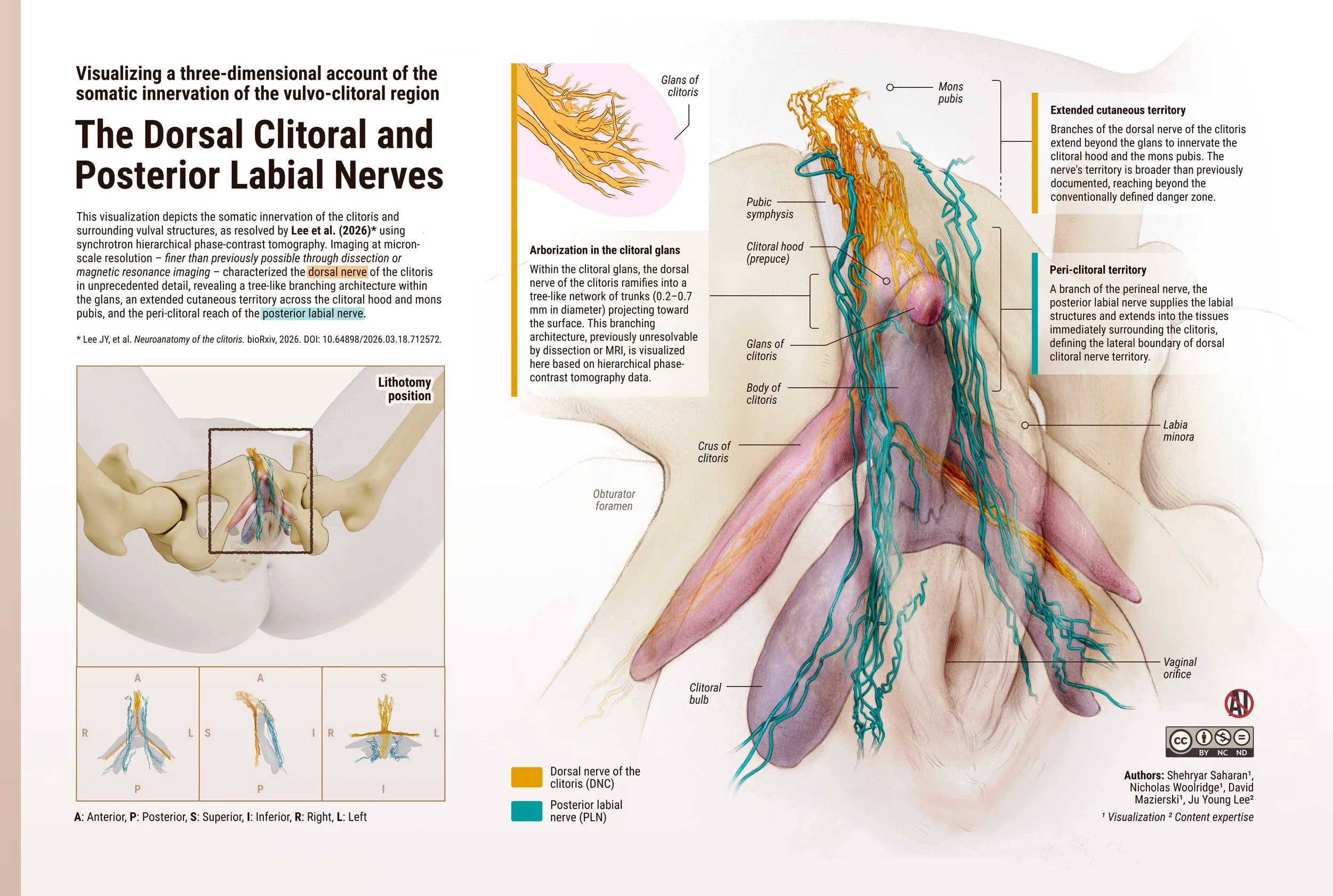
This visualization depicts the somatic innervation of the clitoris and surrounding vulval structures, as resolved by Lee et al. (2026)* using synchrotron hierarchical phase-contrast tomography. Imaging at micron-scale resolution - finer than previously possible through dissection or magnetic resonance imaging - characterized the dorsal nerve of the clitoris in unprecedented detail, revealing a tree-like branching architecture within the glans, an extended cutaneous territory across the clitoral hood and mons pubis, and the peri-clitoral reach of the posterior labial nerve.
Authors: Shehryar Saharan¹, Nicholas Woolridge¹, David Mazierski¹, Ju Young Lee²
¹ Visualization ² Content expertise
* Lee JY, et al. Neuroanatomy of the clitoris. bioRxiv, 2026. DOI: 10.64898/2026.03.18.712572.
This illustration is licensed CC BY-NC-ND 4.0; you're welcome to download and share it for educational and non-commercial purposes, provided it is credited and not altered. A high-resolution file is available below.